




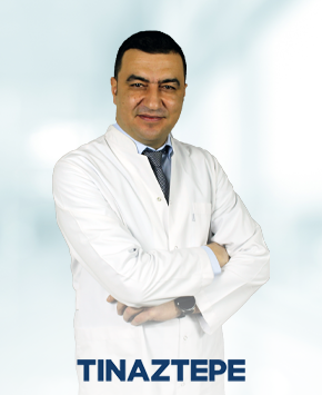
|
Derece
|
Bölüm/Program |
Üniversite |
Yıl |
|
Lise |
Anadolu Lisesi, Almanca |
İzmir Bornova Anadolu Lisesi |
1998 |
|
Lisans |
Tıp Fakültesi |
Celal Bayar Üniversitesi Tıp Fakültesi |
2004 |
|
Tıpta Uzmanlık |
Beyin ve Sinir Cerrahisi |
S.B İzmir Bozyaka Eğitim ve Araştırma Hastanesi |
2010 |
|
Başasistanlık |
Beyin ve Sinir Cerrahisi |
S.B İzmir Bozyaka Eğitim ve Araştırma Hastanesi |
2013 |
|
Doçent |
Beyin ve Sinir Cerrahisi |
İzmir Tınaztepe Üniversitesi |
2022 |
Tıpta Uzmanlık Tezi Başlığı: Laminektomi Defekti Oluşturulmuş Sıçan Modeli Üzerinde Povidon-İodine’in Nörotoksik Etkisinin Araştırılması
Tez Danışmanı: Prof. Dr. Yusuf ERŞAHİN
|
Görev Ünvanı |
Görev Yeri |
Yıl |
|
Asistan Doktor |
İzmir Bozyaka Eğitim ve Araştırma Hastanesi/ İZMİR |
2004-2010 |
|
Uzman Doktor |
İzmir Bozyaka Eğitim ve Araştırma Hastanesi/ İZMİR |
2010-2011 |
|
Uzman Doktor |
Anamur Devlet Hastanesi Anamur/MERSİN |
2011-2012 |
|
Uzman Doktor |
Bakırköy Dr. Sadi Konuk Eğitim ve Araştırma Hastanesi Bakırköy/İSTANBUL |
2012-2013 |
|
Başasistan |
İzmir Bozyaka Eğitim ve Araştırma Hastanesi/ İZMİR |
2013-2021 |
|
Uzman Doktor |
İzmir Tınaztepe Galen Bayraklı Hastanesi |
2021-2022 |
|
Doktor Öğretim Üyesi |
İzmir Tınaztepe Üniversitesi Sağlık Bilimleri Fakültesi |
2022
|
|
Doçent Doktor |
İzmir Tınaztepe Üniversitesi Sağlık Bilimleri Fakültesi / Tınaztepe Galen Bayraklı Hastanesi |
2022-Halen |
Tez Danışmanlığı:
- Uzmanlık Tezi: Deneysel Subaraknoid Kanama Oluşturulmuş Ratlarda Propolis Etkeni Caffeic Acid Phenethyl Esterin Serebral Vasospasm ve Erken Beyin Hasarı Üzerine Etkisi. Dr.Mahmut Necdet Palaz, İzmir, 2016
- Uzmanlık Tezi: Servikal Disk Hernisi Olgularında Anterior Servikal Diskektomi + Füzyon Cerrahisi (ASDF) ve Servikal Disk Protezi (SDP) Ameliyatlarının Postoperatif Dönem Takiplerinde Servikal ROM (Range of motion), Heterotopik Ossifikasyon (HO= Sonuçlarının Retrospektif Olarak İncelenmesi. Dr. Alper Tabanlı, İzmir, 2021
ESERLER
- Uluslararası hakemli dergilerde yayımlanan makaleler:
A1. Akçay E, Erşahin Y, Özer F, Duransoy YK, Çamlar M, Atçı İB, Yağcı A, Özer Ö. “Neurotoxic effect of povidone-iodine on the rat spine using a laminectomy-durotomy model” Childs Nerv Sys. 2012 Dec;28(12):2071-5 doi:10.1007/s00381-012-1885-7
A2. Duransoy YK, Mete M, Akçay E, Selçuki M “Differences in İndividual Susceptibity Affect The Development Of Trigeminal Neuralgia” Neural Regen Research. 2013 8;1337-42 doi:10.3969/j.issn.1673-5374.2013.14.010
A3. Atçı B, Özer FD, Çamlar M, Kocaman Ü, Akçay E, Küpelioğlu A, Erşahin Y, Özer Ö, Karadağ A. “Comparison of Histopathological Outcomes of Three Different Allografts Used for Repairing of The Dural Defect” Journal of Neurological Sciences-Turkish. 2013 30(2);36; 347-55
A4. Özer Ö, Özer FD, Akçay E, Kocaman Ü, Atçı B, Özinel MA, Vardar E, Özdemir N. “A Study on Prophylactic Efficacies of Daptomycin and Vancomycin in a Rat Model of MRSA Infection Secondary to Spinal Implantation” Journal of Neurological Sciences-Turkish. 2013 30(4); 767-75
A5. Aydın M, Akçay E, Yurt A “Is T1-2 Disc Heniation Rare? A Case Report” İnternal Journal of Clinical and Experimental Medicine. 2014 5; 444-46
A6. Aydın M, Gökkara T, Özer FD, Yağcı A, Akçay E, Çamlar M, Aydın T, Erel N “Demineralized Calf Vertebra Model: Can Be Used in Osteoporosis Research?” Journal of Neurological Sciences-Turkish. 2014 31(4);42; 680-86
A7. Engin O, Yildirim M, Kulan A, Dalgic A, Yagci, A., Toptay H & Akcay E. “The free neural grafting for recurrent nerve laceration Experimental study in rabbit.” Annali italiani di chirurgia. 2014 86, 563-569. PMID: 26899952
A8. Yurt A, Seçer M, Aydın M, Akçay E, Ertürk AR, Akkol İ, Yılmaz H, Palaz MN “Surgical management of Juxtafacet cysts in the lumbar spine” İnt J Surg 2016 May;29:9-11 doi: 10.1016/j.ijsu.2016.03.003
A9.Yurt A, Ülgen T, Aydın M, Akçay E, Durmaz O, Benek B, Yılma H, Ertürk AR, Akkol İ, Tabanlı A “Colloid Cysts of the Third Ventricle: 24 Cases Review” Clin Surg. 2018; 3: 2178.
A10. Palaz N, Akçay E “The Impact of Propolis Factor Caffeic Acid Phenethyl-Ester on the Cerebral Vasospasm and Early Brain Damage in the Experimentally Induced Subarachnoid Hemorrhage on Rats” World Neurosurg. 2020 138:e736-e742 doi:10.1016/j.wneu.2020.03.058
A11. Benek HB, Akçay E, Yılmaz H, Yis R, Yurt A “Nocardia cyriacigeorgica brain abscess with Pemphigus vulgaris: first report” 2020 Br J Neurosurg Jan 30:1-2 doi:10.1080/02688697.2020.1716943
A12. Yılmaz H, Akçay E, Benk HB, Yurt A “Guillain-Barre Syndrome After Craniocerebral Gunshot Injury: First Report” 2020, 143:23-25 doi:10.1016/j.wneu.2020.07.103
A13. Akçay E, Yılmaz H, Benek HB, Tabanlı A, Yurt A “Craniopharyngiomas: Analysis of 68 Surgical Cases” Neurological Sciences and Neurophysiology 2020, 37:215-20. doi: 10.4103/NSN.NSN_56_20
A14.Çamlar M, Türk Ç, Arslan FD, Ören MM, Akçay E, Diniz G, Özer F “Does Metformin Protect Against Brain Injury After Subarachnoid Hemorrhage? An Experimental Study” Turkiye Klinikleri J Med Sci.2020;40(4):393-8. doı: 10.5336/medsci.2020-76488
A15. Benek B, Akçay E “Concomitant chronic subdural hematomas and arachnoid cysts in young adults” F1000Research 2021;10.421: 421. doi:10.12688/f1000research.53210.1
A16. Benek B, Akcay E, Yilmaz H, Aydin M, Yurt A “A Comparison of the Surgical Outcomes of Laminoplasty and Laminectomy with Fusion in the Treatment of Multilevel Cervical Spondylotic Myelopathy: A Retrospective Cohort Study” Turkish Neurosurgery 2021; 31(4): 530-537 DOI: 10.5137/1019-5149.JTN.31386-20.2
A17. Yilmaz H, Akcay E, Benek HB “Medulloblastoma in adults: surgical outcomes and survival-a single center analysis of 16 patients.” Turkish neurosurgery,2021; 31(6): 980-985 DOI: 10.5137/1019-5149.JTN.32143-20.2
A18. Benek HB, Akcay E, Yilmaz H, Yurt A “Efficiency of distraction and ligamentotaxis in posterior spinal instrumentation of thoracolumbar retropulsed fractures.” Turkish Neurosurgery 2021;31(6):973-979 DOI: 10.5137/1019-5149.JTN.34860-21.3
A19. Benek B, Yilmaz H, Akçay E, Yurt A “Bilateral Vertebral Artery Occlusion After Traumatic Complete Disruption of the Cervical Spine Associated with Ankylosing Spondylitis.” Turkish Journal of Trauma and Emergency Surgery 2021; 27(6):697-701 DOI: 10.11744/tjtes.2020.6367
- Ulusal hakemli dergilerde yayınlanan makaleler:
B1. Akçay E, Aydın M, Yurt A, Bezircioğlu H, Ak C. “Menengiomu Taklit Eden Soliter Plazmositom Olgusu” İzmir Eğitim Hastanesi Dergisi, Cilt 19,Sayı 4,Ekim-Kasım-Aralık 2015.
B2. Benek HB, Yılmaz H, Akçay E, Yurt A “Atipik Meningiomlar” İzmir Eğitim ve Araştırma Hastanesi Tıp Dergisi (Medical Journal of İzmir Hospital) 2020, 24 (3): 213-218.
B3. Akçay E, Yılmaz H, Benek HB, Yurt A “Beyin Lezyonlarında Nöronavigasyon Cihazı Olarak İntraoperatif Ultrason Kullanımı” İzmir Eğitim ve Araştırma Hastanesi Tıp Dergisi (Medical Journal of İzmir Hospital) 2019, 23 (4): 283-286.
B4. Yılmaz H, Akçay E, Aydın M, Tabanlı A, Kebat T, Ertürl AR, Yurt A “Kistik Menenjiomlar Olgu Serisi” İzmir Eğitim ve Araştırma Hastanesi Tıp Dergisi (Medical Journal of İzmir Hospital) 2019, 23 (4): 343-346.
B5. Yılmaz H, Akçay E Benek HB, Aydın M “Primer Santral Sinir Sistemi Lenfomaları” İzmir Eğitim ve Araştırma Hastanesi Tıp Dergisi (Medical Journal of İzmir Hospital) 2020, 24 (3): 227-231.
B6. Akçay E, Yılmaz H, Benek HB, Tabanlı A, Yurt A “ Surgıcal Resectıon Of Tumors Located In The Insular Regıon” İzmir Eğitim ve Araştırma Hastanesi Tıp Dergisi (Medical Journal of İzmir Hospital) 2020, 24 (2): 161-167.
B7. Benek HB, Akçay E, Yılmaz H, Ülgen T, Tabanlı A, Yurt A “ Serebellopontin Köşe Yerleşimli Tümörlerde Cerrahi Sonuçlarımız” İzmir Eğitim ve Araştırma Hastanesi Tıp Dergisi (Medical Journal of İzmir Hospital) 2021, 25 (1): 57-61.
B8. Benek HB, Akçay E “Spinal Epandimomların Klinik ve Cerrahi Değerlendirilmesi” İzmir Eğitim ve Araştırma Hastanesi Tıp Dergisi (Medical Journal of İzmir Hospital) 2021, 25 (3): 185-189.
- Ulusal bilimsel toplantılarda sunulan ve bildiri kitaplarında basılan bildiriler:
C1. Uçar K, Sayın M, Temiz P, Özer FD, Temiz C, Mete M, Akçay E. “Capae Ekstresi-Allantoin ve Heparinin Lokal Uygulanımı ile Epidural Fibrozisi Önlemedeki Etkinliğinin Araştırılması” Türk Nöroşirürji Derneği 21. Bilimsel Kongresi Özet Kitabı, 130 (2007).
C2. Çamlar M, Özer FD, Duransoy YK, Gökkara T, Yurt A, Akçay E. “Kanamamış İntrakranial Anevrizmaların Cerrahisi” Türk Nöroşirürji Derneği 21. Bilimsel Kongresi Özet Kitabı, 99 (2007).
C3. Erel N, Özer FD, Aydın M, Çamlar M, Akçay E. “Torakal Skolyoz Cerrahisinde Peripediküler Bant Fiksasyonu. Türk Nöroşirürji Derneği 23. Bilimsel Kongresi Özet Kitabı, 48 (2009).
C4. Yurt A, Ertürk AR, Atçı B, Akçay E, Aydın Ö, Çoban G. “3. Ventrikül Kolloid Kistleri” Türk Nöroşirürji Derneği 23. Bilimsel Kongresi Özet Kitabı, 84 (2009).
C5. Özer FD, Aydın M, Akçay E, Yağcı A. “Cauda Equina Paragangliomu” Türk Nöroşirürji Derneği 23. Bilimsel Kongresi Özet Kitabı, 137 (2009).
C6. Yurt A, Selçuki M, Ertürk AR, Yağcı A, Atçı B, Akçay E. “Ekstraventriküler Atipik Kistik Nörositom” Türk Nöroşirürji Derneği 23. Bilimsel Kongresi Özet Kitabı, 178 (2009).
C7. Yurt A, Selçuki M, Ertürk AR, Aydın M, Gökkara T, Akçay E, Kocaman Ü “Hipofiz Tümör İçi Kanamasını Takiben Ortaya Çıkan Akut Serebral Enfarkt” Türk Nöroşirürji Derneği 23. Bilimsel Kongresi Özet Kitabı, 178 (2009).
C8. Yurt A, Akçay E, Aydın M, Aydın Ö, Çoban G “İntraserebral Anevrizma İle Birlikte Olan Anterior Koroidal Arter Anomalileri” Türk Nöroşirürji Derneği 23. Bilimsel Kongresi Özet Kitabı, 187 (2009).
C9. Akçay E, Erşahin Y, Özer FD, Çamlar M, Atçı İB, Yağcı A, Özer Ö “Laminektomi Defekti Oluşturulmuş Sıçan Modeli Üzerinde Povidon-İodine’in Nörotoksik Etkisinin Araştırılması Türk Nöroşirürji Derneği 25. Bilimsel Kongresi Özet Kitabı, (2011).
C10. Özer Ö, Özer FD, Akçay E, Kocaman Ü, Atçı B, Özinel MA, Vardar E, Özdemir N. “A Study on Prophylactic Efficacies of Daptomycin and Vancomycin in a Rat Model of MRSA Infection Secondary to Spinal Implantation” Türk Nöroşirürji Derneği 28. Bilimsel Kongresi Özet Kitabı, 93-4,(2014).
C11. Aydın M, Akkol İ, Akçay E, Bezircioğlu H “C2-C3 Fraktür Dislokasyon Olgusunda Transservikal Paraferengeal Yaklaşımla Anterior Stabilizasyon: Olgu sunumu ve Operasyon Tekniği” Türk Nöroşirürji Derneği 28. Bilimsel Kongresi Özet Kitabı, 209-10(2014).
C12. Akçay E, Aydın M, Yurt A, Bezircioğlu H, Ak C “Menengiomu Taklit Eden Soliter Plasmositom Olgusu” Türk Nöroşirürji Derneği 28. Bilimsel Kongresi Özet Kitabı, 272 (2014).
C13. Aydın M, Akçay E, Yurt A “T1-2 Disk Hernisi Olgu Sunumu” Türk Nöroşirürji Derneği 28. Bilimsel Kongresi Özet Kitabı, 224-5 (2014).
C14. Çoban G, Aydın M, Ak C, Bezircioğlu H, Akçay E “ Tek Seviye Servikal Diskektomi ve Peek Kafes Uygulanan Hastaların Radyolojik ve Klinik Sonuçlarla Değerlendirilmesi” Türk Nöroşirürji Derneği 28. Bilimsel Kongresi Özet Kitabı, 210,(2014).
C15. Ak C, Akkol İ, Akçay E, Aydın M, Bezircioğlu H “ T10-11 Kalsifiye Disk” Türk Nöroşirürji Derneği 28. Bilimsel Kongresi Özet Kitabı, 218,(2014).
C16. Akçay E, Ak C, Palaz MN, Ertürk AR, Akkol İ, Aydın M, Ülgen T, Bülbül Ö, Güler E, Yurt A “Anterior serebral arterin proximal (A1) segmentinin nadir bir varyasyonu. Olgu sunumu” Türk Nöroşirürji Derneği 29. Bilimsel Kongresi Özet Kitabı, 215-6,(2015).
C17. Akçay E, Aydın M, Palaz MN, Ak C, Güler E, Ülgen T, Yurt A “Supratentorial Anaplastik Astroblastom: Olgu Sunumu” Türk Nöroşirürji Derneği 29. Bilimsel Kongresi Özet Kitabı, 256,(2015).
C18. Aydın M, Akçay E, Çoban G, Ülgen T, Kebat T, Güler E, Yurt A “Lateral Ventrikül Hemanjioperisitomu: Olgu Sunumu” Türk Nöroşirürji Derneği 29. Bilimsel Kongresi Özet Kitabı, 143,(2015).
C19. Aydın M, Akçay E, Akkol İ, Yurt A “Lomber Vertebra Metastazları: Retrospektif Değerlendirme Ve Literatüre Uyum” Türk Nöroşirürji Derneği 30. Bilimsel Kongresi Özet Kitabı, 94,(2016).
C20. Aydın M, Durmaz MO, Akçay E, Yurt A “104 Kifoplasti Prosedürünün Erken Post Op Değerlendirilmesi” Türk Nöroşirürji Derneği 30. Bilimsel Kongresi Özet Kitabı, 95,(2016).
C21. Aydın M, Durmaz MO, Akçay E, Akkol İ, Yurt A “Magerl Tip A Kırıklarda Kifoplasti Uygulaması” Türk Nöroşirürji Derneği 30. Bilimsel Kongresi Özet Kitabı, 114,(2016).
C22. Aydın M, Durmaz MO, Akçay E, Palaz MN, Yurt A “Olgu Sunumu: Kliniğimizde 2016 Senesinde Opere Edilen Distal Anterior Serebral Arter Anevrizmalı 78 Y K Hasta” Türk Nöroşirürji Derneği 30. Bilimsel Kongresi Sözlü Sunumlar, 241,(2016).
C23. Aydın M, Durmaz O, Akçay E, Akkol İ, Yurt A “Bozyaka Eğitim ve Araştırma Hastanesinde Opere Edilen Üst Servikal Travmalı Hastaların Retrospektif Analizi” Türk Nöroşirürji Derneği 31. Bilimsel Kongresi Sözlü Sunumlar, 58,(2017).
C24. Durmaz MO, Aydın M, Akçay E, Ertürk AR, Mutlu ŞŞ “Spinal İntradural Tümörler: Retrospektif Değerlendirme Ve Literatüre Uyum” Türk Nöroşirürji Derneği 31. Bilimsel Kongresi Sözlü Sunumlar, 141,(2017).
C25. Aydın M, Tabanlı A, Durmaz O, Akçay E, Yurt A “Tek Merkezli 671 Lomber Disk Herniasyonu Olgusunun Retrospektif Değerlendirilmesi” Türk Nöroşirürji Derneği 31. Bilimsel Kongresi Sözlü Sunumlar, 143,(2017).
C26. Durmaz MO, Yurt A, Akkol İ, Akçay E, Ak C “Vertebral Arter-Posterior İnferior Serebellar Arter Bileşkesinde Tromboze Dev Anevrizma” Türk Nöroşirürji Derneği 31. Bilimsel Kongresi Elektronik Poster Sunumlar, 161-162, (2017).
C27. Durmaz MO, Yurt A, Aydın M, Akçay E, Ak C “Nöropsikolojik Semptomlara Sebep Olan Frontal Konveksite Yerleşimli Araknoid Kist Olgusu” Türk Nöroşirürji Derneği 31. Bilimsel Kongresi Elektronik Poster Sunumlar, 187, (2017).
C28. Durmaz MO, Yurt A, Akçay E, Benek HB, Palaz MN “Post-op Dönemde Aseptik Menekjit Gelişen Epidermoid Kist Olgusu” Türk Nöroşirürji Derneği 31. Bilimsel Kongresi Elektronik Poster Sunumlar, 191, (2017).
C29.Durmaz MO, Yurt A, Aydın M, Akçay E, Ülgen T “Sağ Hemiparezi ve Disfazi İle Başvuran Dev Atipik Menenjiom Olgusu” Türk Nöroşirürji Derneği 31. Bilimsel Kongresi Elektronik Poster Sunumlar, 194-195, (2017).
C30. Aydın M, Durmaz MO, Akçay E, Akkol İ, Benek HB, Ülgen T, Tabanlı A, Yurt A “Alt Servıkal Travma Nedenıyle Opere Edılen 25 Hastanın Değerlendırılmesı: En Az İki Yıl Takıp” Türk Nöroşirürji Derneği 32. Bilimsel Kongresi Sözlü Sunumlar, 116,(2018).
C31. Aydın M, Benek HB, Akçay E, Durmaz MO, Akkol İ, Yurt A “Servıkal Pedıkül Vıdası Deneyımlerımız” Türk Nöroşirürji Derneği 32. Bilimsel Kongresi Sözlü Sunumlar, 119,(2018).
C32. Aydın M, Durmaz MO, Akçay E, Göçmen Mas NN, Yurt A “Ika Lacerum Segmentı, For. Spınosum ve For. Ovale Arasındakı Anatomık Ilışkı: Anatomık Çalışma” Türk Nöroşirürji Derneği 32. Bilimsel Kongresi Sözlü Sunumlar, 141, (2018).
C33. Benek HB, Akçay E, Aydın M, Ülgen T, Tabanlı A, Yurt A “Denovo Torakal Schwannom: Total Rezeksiyondan 4 Yıl Sonra Farklı Spinal Seviyede Yeni Gelişmiş Schwannom Olgusu” Türk Nöroşirürji Derneği 32. Bilimsel Kongresi Elektronik Poster Sunumlar, 206, (2018).
C34. Akçay E, Benek HB, Aydın M, Tabanlı A, Yurt A “İntrakranial Meningiomlar: 98 Olgunun Who 2016 Evreleme Sistemi İle Retrospektif Analizi” Türk Nöroşirürji Derneği 32. Bilimsel Kongresi Elektronik Poster Sunumlar, 209, (2018).
C35. Akçay E, Benek HB, Aydın M, Durmaz MO, Akkol İ, Yurt A, Tabanlı A “Sakral Kordoma Öntanılı, İntraosseöz Dev İnvaziv Schwannom Olgusu” Türk Nöroşirürji Derneği 32. Bilimsel Kongresi Elektronik Poster Sunumlar, 316, (2018).
C36 Akçay E, Yılmaz H, Benek HB “Spinal Tümörlerde Cerrahi Tedavi Sonuçlarımız” Türk Nöroşirürfi Derneği Spinal ve Periferik Sinir Cerrahisi Eğitim Öğretim Grubu Yaşlanan Omurga Sempozyumu Türk Nöroşir Derg 29(Ek sayı 1),s:58, 2019
C37. Yılmaz H, Akçay E, Benek HB “Primer Santral Sinir Sistemi Lenfoması: Olgu Sunumu” 7. Bozyaka Hematoloji Sempozyumu Özet Kitabı, 30, 2019
C38 Benek B, Yılmaz H, Akçay E “Glioblastoma Multiforme Tanılı 87 Hastanın Retrospektif Analizi” NOVA 2019 Sempozyumu | Sözlü Sunumlar SB-1, Türk Nöroşir Derg 30(Ek sayı 1), s: 24, 2020
C39. Benek B, Yılmaz H, Akçay E “Tıp 2 Odontoıd Kırıkları Tedavısınde Uygulanan Anterıor Odontoıd Vıdalama Sonuçlarımız” Spinal Travmalar Sanal Sempozyumu Bildiri Özet Kitapçığı SB-5 (5795),s:32, 2020
C40. Benek HB, Akçay E, Yılmaz H “Uzak Lateral Yerleşimli Lomber Disk Hernileri Cerrahi Sonuçlarımızın Değerlendirilmesi “ Spinal Kökenli Bel ve Bacak Ağrısı Sempozyumu Bildiri Özet Kitabı SB-11, s: 50; 2021
C41. Tabanlı A, Akçay E, Benek HB, Yılmaz H, Boloğur O, Kayıkçı E, Yurt A “2000-2020 Yılları Arasında Yapılan Mikrocerrai Anevrizma Teadvilerinin Değerlendirilmesi” Türk Nöroşirürji Derneği 34. Bilimsel Kongresi, Sözlü Sunumlar SS-150, Türk Nöroşir Derg 31(Ek Sayı-1); s:129, 2021
C42. Tabanlı A, Şensoy T, Boloğur O, Kayıkçı E, Akçay E, Benek HB, Yılmaz H, Yurt A, Ülgen T “2015-2019 Yılları Arasında Yapılan Hipofiz Adenomlarının Transsfenoidal veTranskranial Tedavilerinin Değerlendirilmesi” Türk Nöroşirürji Derneği 34. Bilimsel Kongresi, Sözlü Sunumlar SS-150, Türk Nöroşir Derg 31(Ek Sayı-1); s:129-130, 2021
D. Uluslararası bilimsel toplantılarda sunulan ve bildiri kitaplarında basılan bildiriler:
D1. Benek B, Akçay E, Aydın M, Durmaz O, Yurt A “Spontaneous Complete Resolution of Two Sequestrated Lumbar Disc Herniations at Different Levels and Time Periods in the Same Patient” WFNS-2017 XVI. World Congress of Neurosurgery, Abstract book OP-SP.24-08, pp280, 2017
D2. Yurt A, Akçay E, Aydın M, Ülgen T, Durmaz O, Benek HB, Akkol İ, Ertürk AR, Tabanlı A, Ak C, Mutlu Ş “Surgical Results of 500 Cases of Intracranial Aneurysms WFNS-2017 XVI. World Congress of Neurosurgery, Abstract book EP-0094, pp322, 2017
D3. Benek B, Aydın M, Akçay E, Ülgen T, Akkol İ, Yurt A “Aggresive Vertebral Hemangioma of the Thoracic Spine” WFNS-2017 XVI. World Congress of Neurosurgery, Abstract book EP-0377, pp408-409, 2017
D4. Durmaz MO, Aydın M, Ertürk AR, Akçay E, Ülgen T, Tabanlı A “Cerebellar Hemangiopericytoma: A Rare Case Report and Review of the Literature” WFNS-2017 XVI. World Congress of Neurosurgery, Abstract book EP-0677, pp499, 2017
D2. Akçay E, Benek HB “Unilateral Percutaneous Kyphoplasty in Thoracolumbar Compression Fractures” 5. Uluslararası Hipokrat Tıp ve Sağlık Bilimleri Kongresi Bildiri Özet Kitabı SS247, s: 899, 2020
E. Yazılan ulusal/uluslararası kitaplar veya kitaplardaki bölümler:
E1. Omurilik ve Omurga tümörleri Editör:Dalbayrak Sedat, Yaman Onur, Kaptanoğlu Erkan, Şimşek Serkan, Ateş özkan, Dalgıç Ali, Bölüm adı: “Spinal tümör cerrahisinde nöromonitörizasyon” Akçay Emrah, Dalgıç Ali Türk Nöroşirürji Derneği SPSCG Yayınları No:16; s:364-376, 2014, Ankara ISBN:978-605-4149-16-2 Türkçe(Bilimsel kitap)
E2. Spinal deformiteler Editör:Dalbayrak Sedat, Yaman Onur, Kaptanoğlu Erkan, Şimşek Serkan, Ateş Özkan, Dalgıç Ali, Bölüm adı: “İdiopatik skolyoz cerrahisinde erken ve geç sorunlar” Aydın Murat, Akçay Emrah, Türk Nöroşirurji Derneği SPSCG Yayınları No:18, 2015, Ankara ISBN:978-605-4149-16-2, Türkçe(Bilimsel Kitap)
E3. Spinal deformiteler Editör:Dalbayrak Sedat, Yaman Onur, Kaptanoğlu Erkan, Şimşek Serkan, Ateş Özkan, Dalgıç Ali, Bölüm adı: “Hemivertebrektomi: Endikasyon ve Teknik” Akçay Emrah, Aydın Murat, Sedat Dalbayrak Türk Nöroşirurji Derneği SPSCG Yayınları No:18, 2015, Ankara, ISBN:978-605-4149-16-2, Türkçe(Bilimsel Kitap)
E4. Nöroşirürji Yeterlilik Sınavına Hazırlık Editör: Biçeroğlu Hüseyin, Hancı Murat, Bölüm adı: “Sakral ve lumbosakral bileşke lezyonlarına yaklaşım” Akçay E, US Akademi Yayınları, cilt:2 sayfa 375-388, 2016, İzmir ISBN:978-605-9358-05-7, 978-605-9358-04-0, 978-605-9358-03-3 (TK) , (Türkçe bilimsel kitap)
E5. Nöroşirürjinin Temelleri Editör: Naderi S, Karagöz Güzey F, Tuğcu, Bölüm Adı: “Epilepsi Patofizyolojisi, Epilepsi Çeşitleri, Medikal Tedavi, Epilepsi Cerrahisine Uygun Hasta Seçimi Ve Ameliyat Öncesi Değerlendirme” Akçay E, US Akademi Yayınları sayfa 531-536, 2020, İzmir ISBN: 978-605-9358-81-1, Türkçe (Bilimsel Kitap)
F.Katılınan kurs,sempozyum ve kongreler:
F1. TND XXI. Bilimsel Kongresi 20-24 Nisan 2007, Antalya
F2. TND Spinal ve Periferik Sinir Cerrahisi Grubu “Spinal Cerrahi Yaz Okulu 1. Kurs” 26-29 Haziran 2008, Denizli
F3. TND XXIII. Bilimsel Kongresi 17-21 Nisan 2009, Girne,KKTC
F4. Dokuz Eylül Üniversitesi Tıp Fakültesi “Deney Hayvanları Kullanım Sertifikası” Kursu Kasım 2009, İzmir
F5. İzmir İl Sağlık Müdürlüğü “Organ Temininde Yeni Adımlar: Yoğun Bakım Eğitimi” kurs programı Nisan 2010, İzmir
F6. TND XXIV. Bilimsel Kongresi 14-18 Mayıs 2010, Antalya
F7. TND XXV. Bilimsel Kongresi 14-18 Mayıs 2011, Antalya
F8. TND Spinal ve Periferik Sinir Cerrahisi Grubu “Omurga Cerrahisinde Olgu Çözümlemeleri” Sempozyumu 26-29 Eylül 2013, Çeşme
F9. TND XXVIII. Bilimsel Kongresi 3-7 Nisan 2014, Antalya
F10. TND Spinal ve Periferik Sinir Cerrahisi Grubu “Mehmet Zileli İleri Spinal Cerrahi Kursu” 5-7 Aralık 2014, İzmir
F11. TND XXIX. Bilimsel Kongresi 17-21 Nisan 2015, Antalya
F12. TND Spinal ve Periferik Sinir Cerrahisi Grubu “Spinal Deformiteler” Sempozyumu 29 Ekim-1 Kasım 2015, Antalya
F13. İzmir Organ Doku Nakli Bölge Koordinasyon Merkezi “Beyin Ölümü Tanısı ve Donör Bakımı Eğitimi” 14 Aralık 2015, İzmir
F14. TND XXX. Bilimsel Kongresi 8-12 Nisan 2016, Antalya
F15. TND Spinal ve Periferik Sinir Cerrahisi Grubu “Lomber Dejeneratif Omurga Sonbahar Sempozyumu” 20-23 Ekim 2016, Antalya
F16. T.C. S.B. İzmir İli Güney Bölgesi Kamu Hastaneler Birliği Genel Sekreterliği “Organ Nakli Sempozyumu” 1 Kasım 2016, İzmir
F17. TND Spinal ve Periferik Sinir Cerrahisi Grubu “Servikal ve Torakal Dejeneratif Omurga Sempozyumu” 21-24 Eylül 2017, Bodrum
F18. İzmir Bozyaka Eğitim ve Araştırma Hastanesi “Gamma Knife Radyocerrahisi Güncel Gelişmeler” Eğitim Semineri, 5 Aralık 2017, İzmir
F19. TND XXXI. Bilimsel Kongresi 29 Mart-2 Nisan 2017, Antalya
F20. Sağlık Bilimleri Üniversitesi Beyin ve Sinir Cerrahisi Anabilim Dalı “Nörotravmatoloji Toplantısı” 17 Şubat 2018, İstanbul
F21. TND XXXII. Bilimsel Kongresi 20-24 Nisan 2018, Antalya
F22. TND XXXIII. Bilimsel Kongresi 11-14 Nisan 2019, Antalya
F23. TND Spinal ve Periferik Sinir Cerrahisi Grubu “Yaşlanan Omurga Sempozyumu” 31 Ekim- 3 Kasım 2019, Antalya
F24. 2021World Spina Column Society “Virtual World Spine 2020” 29-30 Ağustos 2020
F25. TND Spinal ve Periferik Sinir Cerrahisi Grubu “Spinal Travmalar Sanal Sempozyumu” 20 Ekim 2020
F26. Sinir Sistemi Cerrahisi Derneği “ Vasküler Nöroşirürji e-Sempozyumu” 6 Şubat 2021
F27. TND XXXIV. Bilimsel Kongresi 30Eylül-3 Ekim 2021, çevrimiçi
DOÇ. DR. EMRAH AKÇAY
GÖREV ALDIĞI BÖLÜMLER
BEYİN VE SİNİR CERRAHİSİ


THE EFFECTS OF PREOPERATIVE CARBOHYDRATE LOADING ON POSTOPERATIVE OUTCOMES IN NEUROSURGERY: A SYSTEMATIC REVIEW AND META-ANALYSIS
By
Sanli, D (Sanli, Deniz) [1] ; Kundakçi, GA (Kundakci, Gamze Agartioglu) [2] ; Katircioglu, K (Katircioglu, Kaan) [3] ; Yurt, A (Yurt, Alaettin) [4] ; Akçay, E (Akcay, Emrah) [4]
(provided by Clarivate)
Source
Volume
25
Issue
1
DOI
10.1186/s12871-025-03468-2
Article Number
575
Published
NOV 19 2025
Indexed
2025-11-26
Document Type
Review
Abstract
Background Preoperative carbohydrate loading is a crucial component of ERAS protocols. However, its effectiveness has not previously been evaluated in neurosurgery. This systematic review and meta-analysis aimed to assess the postoperative effectiveness of preoperative carbohydrate loading in neurosurgery. Methods The EBSCOhost, PubMed, Scopus, Ovid LWW Total Access, Web of Science, and Cochrane Central Register of Controlled Trials databases were searched up to November 2024. Randomized and nonrandomized controlled trials involving patients aged 18 and above who underwent any type of elective neurosurgery and received any form of preoperative oral carbohydrate beverage were included. The primary outcomes were total and postoperative length of hospital stay (LOS). The secondary outcomes were 30-day readmission, total cost of hospitalization, ambulation on postoperative day (POD) 1, oral intake time, pain, postoperative nausea and vomiting (PONV), and complications. The methodological quality and risk of bias of the selected studies were assessed. Review Manager 5.4 software and Comprehensive Meta-Analysis v4 software were used for the statistical analyses. Results A total of nine studies involving 1,299 patients were included in the assessment of total hospital LOS, and six studies involving 761 patients were included in the examination of postoperative hospital LOS. The total hospital LOS (MD = -1.75 days, 95% CI: -2.78 to -0.73, p = 0.0008) and postoperative hospital LOS (MD = -3.40 days, 95% CI: -4.80 to -2.00, p < 0.00001) were significantly shorter in the intervention group. In addition, the intervention group had a significantly higher rate of ambulation on POD 1 (OR = 12.84, 95% CI: 2.85-57.77, p = 0.0009), whereas the rates of pain (OR = 0.18, 95% CI: 0.13-0.26, p < 0.00001) and PONV (OR = 0.37, 95% CI: 0.25-0.53, p < 0.00001) decreased. The results regarding the total cost of hospitalization, oral intake time, and respiratory system complications should be interpreted with caution. Conclusion Preoperative carbohydrate loading is effective in improving postoperative patient outcomes during neurosurgery. It is recommended that institutions support preoperative carbohydrate loading by establishing guideline-based written protocols and integrating it with other evidence-based strategies to enhance surgical outcomes.Trial registrationRegistration number International prospective register of systematic reviews (PROSPERO): CRD42024577131.
Keywords
Author Keywords
Neurosurgery Postoperative outcomes Preoperative carbohydrate loading Meta-analysis Systematic review
Surgical outcomes of tethered cord syndrome in patients with normal conus medullaris and filum terminale without urologic symptoms
By
Tabanli, A (Tabanli, Alper) [1] ; Akcay, E (Akcay, Emrah) [1] ; Yilmaz, H (Yilmaz, Hakan) [2] ; Ozdemir, S (Ozdemir, Seymen) [3] ; Mete, M (Mete, Mesut) [4] ; Selcuki, M (Selcuki, Mehmet) [1]
(provided by Clarivate)
Source
Volume
41
Issue
1
DOI
10.1007/s00381-024-06713-0
Article Number
47
Published
DEC 2025
Indexed
2024-12-25
Document Type
Article
Jump to
Abstract
PurposeTethered cord syndrome (TCS) typically presents with urologic symptoms and abnormal imaging findings. However, some patients present with normal conus medullaris level and filum terminale appearance on MRI. This research seeks to assess the intended surgical results in this particular group of TCS patients who do not present with urologic complaints, under the premise that the surgical approach goes a long way in preventing the onset of urologic abnormalities.MethodsThis retrospective study included 59 operated patients with tethered cord syndrome who had a normal level terminating conus medullaris and a normal looking filum terminale without urologic symptoms. Of these patients, 38 were female and 21 were male. All patients underwent somatosensory-evoked potentials (SSEPs), and magnetic resonance imaging (MRI). The surgical technique used was flavotomy, which involves cutting the filum terminale without performing a laminectomy.ResultsThe study population had a mean age of 22.5 years (SD = 13.2). During the mean postoperative follow-up period of 2.5 years, none of the patients developed urinary incontinence. Preoperative SSEP abnormalities included conduction block in 39 patients (66.1%), low amplitude in 12 patients (20.3%), and delayed N22 wave latency in 8 patients (13.5%). The surgical procedures were completed without morbidity or mortality, and all patients showed significant postoperative improvement in SSEP parameters.ConclusionOur results indicate that even though the filum terminale might have a normal looking MRI, TCS can also occur due to some potential microscopic or structural abnormality. The study proves SSEP to be useful in TCS diagnosis and it also proposes that if surgery is done early before any urologic complaints arise, chances of their onset will be minimized. Such findings support the view that surgical measures should be entertained in symptomatic patients with abnormal SSEP but normal MRI.
Author Keywords
Tethered cord syndrome Conus medullaris Filum terminale Somatosensory-evoked potentials
ChatGPT-4o's Performance in Brain Tumor Diagnosis and MRI Findings: A Comparative Analysis with Radiologists
By
Ozenbas, C (Ozenbas, Cemre) [1] ; Engin, D (Engin, Duygu) [2] ; Altinok, T (Altinok, Tayfun) [1] ; Akcay, E (Akcay, Emrah) [3] ; Aktas, U (Aktas, Ulas) [4] ; Tabanli, A (Tabanli, Alper) [4]
(provided by Clarivate)
Source
Volume
32
Issue
6
Page
3608-3617
DOI
10.1016/j.acra.2025.01.033
Published
JUN 2025
Early Access
MAY 2025
Indexed
2025-06-17
Document Type
Article
Jump to
Abstract
Rationale and objectives: To evaluate the accuracy of ChatGPT-4o in identifying magnetic resonance imaging (MRI) findings and diagnosing brain tumors by comparing its performance with that of experienced radiologists. Materials and methods: This retrospective study included 46 patients with pathologically confirmed brain tumors who underwent preoperative MRI between January 2021 and October 2024. Two experienced radiologists and ChatGPT 4o independently evaluated the anonymized MRI images. Eight questions focusing on MRI sequences, lesion characteristics, and diagnoses were answered. ChatGPT-4o's responses were compared to those of the radiologists and the pathology outcomes. Statistical analyses were performed, which included accuracy, sensitivity, specificity, and the McNemar test, with p<0.05 considered to indicate a statistically significant difference. Results: ChatGPT-4o successfully identified 44 of the 46 (95.7%) lesions; it achieved 88.3% accuracy in identifying MRI sequences, 81% in perilesional edema, 79.5% in signal characteristics, and 82.2% in contrast enhancement. However, its accuracy in localizing lesions was 53.6% and that in distinguishing extra-axial from intra-axial lesions was 26.3%. As such, ChatGPT-4o achieved success rates of 56.8% and 29.5% for differential diagnoses and most likely diagnoses when compared to 93.2-90.9% and 70.5-65.9% for radiologists, respectively (p<0.005). Conclusion: ChatGPT-4o demonstrated high accuracy in identifying certain MRI features but underperformed in diagnostic tasks in comparison with the radiologists. Despite its current limitations, future updates and advancements have the potential to enable large language models to facilitate diagnosis and offer a reliable second opinion to radiologists.
Keywords
Author Keywords
Artificial intelligence ChatGPT-4oBrain tumor MRI
IS UNILATERAL EXTENDED PTERIONAL CRANIOTOMY ADEQUATE INSTEAD OF BICORONAL (BIFRONTAL) CRANIOTOMY IN LARGE OR GIANT OLFACTORY GROOVE MENINGIOMAS?
By
Yilmaz, H (Yilmaz, Hakan) [1] ; Akcay, E (Akcay, Emrah) [1] ; Tabanli, A (Tabanli, Alper) [1] ; Bologur, O (Bologur, Onur) [1] ; Cafer, AK (Cafer, A. K.) [1] ; Benek, HB (Benek, Huseyin Berk) [1] ; Yurt, A (Yurt, Alaettin) [1]
(provided by Clarivate)
Source
Volume
35
Issue
1
Page
56-61
DOI
10.5137/1019-5149.JTN.46246-24.3
Published
2025
Indexed
2025-02-13
Document Type
Article
Abstract
AIM: To evaluate the radiological characteristics, clinical features,and surgical outcomes of bicoronal incision and bifrontal craniotomy for olfactory groove meningiomas (OGMs). MATERIAL and METHODS: This was a retrospective review of 16 patients (nine male and seven female) with large and giant OGMs operated through unilateral extended pterional craniotomy between 2010 and 2022. The radiological characteristics, clinical features,and surgical outcomes were examined. RESULTS: All patients underwent surgical resection via a unilateral extended pterional approach.The mean age of patients was 62.1 years. The most common presenting symptoms were altered consciousness, seizures, headache,and anosmia. Ten (62.5%) and 6 (37.5%) patients had large (4-6 cm) and giant (>6 cm) OGMs, respectively. The mean tumor diameter was 6.3 cm (range:4-9). Simpson Grade2 resection was achieved in all 16 patients. CONCLUSION: Unilateral extended pterional craniotomy offers a safe and effective alternative to the bilateral coronal approach for large and giant OGMs, minimizing risks of frontal lobe retraction, brain edema, and venous infarction. This approach allows for total resection with very low morbidity and mortality rates, making it a viable surgical approach for these complex tumors.
Author Keywords
Giant Meningioma Olfactory groove Pterional craniotomy
Comparison of the Outcomes of Anterior Cervical Discectomy and Fusion and Cervical Disc Replacement for Cervical Disc Disease
By
Tabanli, A (Tabanli, Alper) [1] ; Akcay, E (Akcay, Emrah) [1] ; Yilmaz, H (Yilmaz, Hakan) [1] ; Ak, C (Ak, Cafer) [1] ; Bologur, O (Bologur, Onur) [1] ; Kayikci, E (Kayikci, Engin) [1]
(provided by Clarivate)
Source
JCPSP-JOURNAL OF THE COLLEGE OF PHYSICIANS AND SURGEONS PAKISTAN
Volume
34
Issue
5
Page
551-555
DOI
10.29271/jcpsp.2024.05.551
Published
MAY 2024
Indexed
2024-06-29
Document Type
Article
Abstract
Objective: To compare the radiological outcome and development of heterotopic ossification (HO) following single-segment anterior cervical discectomy and fusion (ACDF) and cervical disc replacement (CDR) for cervical disc herniation and evaluate their impact on surgical success.
Study Design: Descriptive comparative study.
Place and Duration of the Study: Neurosurgery Department at Bozyaka Education and Research Hospital, Izmir, Turkiye, between January 2020 and June 2022.
Methodology: Patients aged 18-70 years with radicular neck pain unresponsive to conventional medical treatment and an MRI-confirmed diagnosis were included. Patients with osteoporosis (OP) were excluded. Patients were randomised into two treatment groups (ACDF and CDR) and stratified by age and symptom severity. Radiographic assessments and HO classification according to McAfee were performed.
Results: Among the included patients, 56 underwent ACDF and 45 underwent CDR. The mean patient age was 48.29 +/- 9.530 and 41.84 +/- 7.239 years in the ACDF and CDR groups, respectively (p <0.001). The postoperative disc height increased in both groups. The T1 slope was significantly higher preoperatively and in the early postoperative period in the CDR group than in the ACDF group (p = 0.001). HO was graded as 1, 2, 3, and 4 in 28 (27.7%), 6 (5.9%), 7 (6.9%), and 4 (3%) patients, respectively.
Conclusion: ACDF and CDR provided similar improvements in radiological measurements and pain relief. Although both procedures significantly enhanced the patient's quality of life and disability scores, HO was more prevalent following CDR during long-term follow-up.
Keywords
Author Keywords
Cervical disc replacement Anterior cervical discectomy and fusion Spinal surgery techniques Heterotopic ossification
Incidence of New Fractures in Patients Treated with Kyphoplasty/Vertebroplasty or Conservative Methods
By
Tabanli, A (Tabanli, Alper) [1] ; Yilmaz, H (Yilmaz, Hakan) [2] ; Benek, HB (Benek, Huseyin Berk) [2] ; Ercan, MA (Ercan, Mehmet Akif) [2] ; Ozgenc, G (Ozgenc, Gulsen) [2] ; Cafer, AK (Cafer, A. K.) [2] ; Bologur, O (Bologur, Onur) [2] ; Akcay, E (Akcay, Emrah) [1] ; Yurt, A (Yurt, Alaettin) [1]
(provided by Clarivate)
Source
Volume
61
Issue
11
DOI
10.3390/medicina61112012
Article Number
2012
Published
NOV 11 2025
Indexed
2025-12-03
Document Type
Article
Jump to
Abstract
Background and Objectives: Osteoporotic vertebral compression fractures (OVCFs) are a common cause of morbidity in the elderly, often resulting in pain, reduced mobility, and diminished quality of life. Treatment options include conservative management, vertebroplasty (VP), and kyphoplasty (KP). This study aimed to compare the clinical outcomes and complication rates of patients treated with kyphoplasty/vertebroplasty versus conservative methods and identify risk factors associated with the development of new fractures. Materials and Methods: This retrospective cohort study included patients diagnosed with OVCFs who were treated either surgically (KP/VP) or conservatively between January 2020 and January 2025. Inclusion criteria encompassed vertebral height loss on CT, STIR hyperintensity on MRI, and a T-score below -2.5. Patients were followed for at least one year. Clinical evaluations included pain scores (VAS), functional status (ODI), and quality of life assessments. Complications and new fracture rates were recorded. Logistic regression analysis was performed to identify risk factors influencing fracture recurrence. Results: A total of 132 patients were analyzed: 65 in the KP/VP group and 67 in the conservative treatment group. The KP/VP group achieved better postoperative pain results (3.2 +/- 1.0 vs. 4.0 +/- 1.2) with a significant difference of -0.8 (95% CI: -1.2 to -0.4, p = 0.032) and better mobility results (ODI: 4.5 +/- 0.8 vs. 3.9 +/- 0.9) with a significant difference of 0.6 (95% CI: 0.3-0.9, p = 0.047) and improved quality of life scores (6.7 +/- 1.1 vs. 5.9 +/- 1.3) with a significant difference of 0.8 (95% CI: 0.4-1.2, p = 0.041). The incidence of new fractures was similar between groups (15.4% vs. 17.9%, p = 0.678). Overall complication rates were 7.7% in the KP/VP group versus 11.9% in the conservative group (p = 0.435). The results from logistic regression analysis showed that age (adjusted OR: 2.48, 95% CI: 1.20-5.13), low bone mineral density (adjusted OR: 0.31, 95% CI: 0.15-0.63), and cement leakage (adjusted OR: 3.10, 95% CI: 1.21-7.99) were identified as risk factors for new fractures. The study found that outdoor activity (adjusted OR: 0.38, 95% CI: 0.20-0.73) and anti-osteoporosis treatment (adjusted OR: 0.17, 95% CI: 0.04-0.79) acted as protective factors against new fractures. The KP/VP group required half the time to recover from their injuries because they used their braces for 3.0 +/- 0.5 months instead of 6.0 +/- 1.0 months (p < 0.001). Conclusions: Kyphoplasty and vertebroplasty were more effective than conservative treatment in improving pain, mobility, and quality of life in patients with OVCFs. Although the incidence of new fractures did not differ significantly between groups, surgical treatment demonstrated lower complication rates and significantly faster recovery, as evidenced by reduced brace use duration. These findings support the use of KP/VP as a viable option for managing OVCFs in appropriately selected patients.
Author Keywords
osteoporotic vertebral compression fracture kyphoplasty vertebroplasty
Trigeminal neuralgia secondary to compression of giant quadrigeminal cistern arachnoid cyst in an adult: Case report and review of the literature
By
Benek, HB (Benek, Huseyin Berk) [1] ; Yilmaz, H (Yilmaz, Hakan) [1] ; Tabanli, A (Tabanli, Alper) [1] ; Akçay, E (Akcay, Emrah) [1] ; Yurt, A (Yurt, Alaattin) [1]
(provided by Clarivate)
Source
AGRI-THE JOURNAL OF THE TURKISH SOCIETY OF ALGOLOGY
Volume
37
Issue
3
Page
175-178
DOI
10.14744/agri.2023.70845
Published
JUL 2025
Indexed
2025-10-23
Document Type
Review
Abstract
A quadrigeminal cistern arachnoid cyst is an extremely rare cause of trigeminal neuralgia in adulthood.This report presents the fourth case in the literature of trigeminal neuralgia caused by a giant quadrigeminal cistern arachnoid cyst. A 52-year-old woman presented with a 3-month history of cerebellar ataxia and vertigo. She had a 2-year history of trigeminal neuralgia that was unresponsive to medical therapy. Neuroimaging studies revealed a cystic lesion in the quadrigeminal cistern associated with hydrocephalic enlargement. Using an infratentorial supracerebellar approach, neuroendoscope-assisted decompression and fenestration of the cyst were performed. Postoperatively, the patient was pain-free and able to walk unaided without symptoms of ataxia.
Keywords
Author Keywords
Arachnoid cyst ataxia hydrocephalus quadrigeminal cistern trigeminal neuralgia
Hemorrhagic Risk in Vestibular Schwannoma Surgeries: Insights and Implications
By
Tabanli, A (Tabanli, Alper) [1] ; Yilmaz, H (Yilmaz, Hakan) [2] ; Akçay, E (Akcay, Emrah) [1] ; Benek, HB (Benek, Huseyin Berk) [2] ; Atci, IB (Atci, Ibrahim Burak) [3] ; Mete, M (Mete, Mesut) [4]
(provided by Clarivate)
Source
Volume
30
DOI
10.12659/MSM.946583
Article Number
e946583
Published
DEC 30 2024
Indexed
2025-01-13
Document Type
Article
Abstract
Background: Vestibular schwannoma is a slow-growing benign tumor arising from the 8th cranial nerve. It can originate in the cerebellopontine angle (CPA). This retrospective study aimed to investigate the factors associated with outcomes following surgical resection of vestibular schwannoma in the CPA in 30 patients at a single center in Turkey, focusing on postoperative intratumoral hemorrhage. Material/Methods: Thirty patients (mean age 42.8 years, range 17-81) underwent vestibular schwannoma surgery via a lateral suboccipital retrosigmoid approach. Patients were categorized as 'less bleeding' (n=15) or 'more bleeding' (n=15) based on the intraoperative nature of the tumor. Demographic characteristics, tumor size, extent of resection, postoperative intratumor bleeding rates, morbidity, and mortality were evaluated. Results: Mean tumor size was significantly larger in highly hemorrhagic tumors (3.8 cm, range 2.1-5 cm) compared with less hemorrhagic tumors (2.1 cm, range 1.8-3 cm) (P<0.001). Total resection was achieved in 60% of patients with highly hemorrhagic tumors >3 cm and chronic diseases, compared with 80% in less hemorrhagic tumors (P=0.02). Postoperative intratumoral hemorrhage occurred in 83.3% of subtotal resections in highly hemorrhagic tumors, versus 6.7% in less hemorrhagic tumors (P<0.001). Conclusions: Larger vestibular schwannoma size is associated with increased hemorrhagic nature, complicating total resection. Subtotal resection in hemorrhagic tumors significantly increases the risk of postoperative bleeding and edema. When possible, total removal should be attempted to minimize complications. In cases requiring subtotal excision, careful postoperative management of coagulation and blood pressure is crucial.
Author Keywords
Edema Hemorrhage Schwann Cells
Investigation of the Effects of Quercetin on Early Brain Injury and Vasospasm in an Experimental Model of Subarachnoid Hemorrhage
By
Kayikci, E (Kayikci, Engin) [1] ; Yilmaz, H (Yilmaz, Hakan) [1] ; Benek, HB (Benek, Huseyin Berk) [1] ; Akcay, E (Akcay, Emrah) [1] ; Tabanli, A (Tabanli, Alper) [1] ; Bologur, O (Bologur, Onur) [1] ; Ak, C (Ak, Cafer) ; Soylu, E (Soylu, Emrah) [2] ; Kebat, T (Kebat, Tulu) [3] ; Bozkaya, G (Bozkaya, Giray) [4] ;
(provided by Clarivate)
Source
Volume
35
Issue
6
Page
891-898
DOI
10.5137/1019-5149.JTN.48407-25.2
Published
2025
Indexed
2025-12-06
Document Type
Article
Abstract
AIM: To investigate the therapeutic efficacy of quercetin in early brain injury (EBI) and vasospasm resulting from experimental subarachnoid hemorrhage (SAH). MATERIAL and METHODS: The rats were assigned into five groups, as follows; Group A: rats did not undergo SAH induction, Group B: rats underwent SAH induction, but received no treatment, Group C: rats underwent SAH induction and received dimethyl sulfoxide (DMSO) intraperitoneally, Group D: rats underwent SAH induction and followed by the intraperitoneal administration of 10 mg quercetin, Group E: rats underwent SAH induction, followed by the intraperitoneal administration of 50 mg quercetin. After the procedure, each group of rats received DMSO, 10 mg/kg quercetin, or 50 mg/kg quercetin intraperitoneally at 30 minutes, 12 hours, and 24 hours, according to their respective categories. The oxidative stress index (OSI) was biochemically measured using the total oxidant status and total antioxidant status. The serum caspase-3, glutathione peroxidase-1 (GPX), and malondialdehyde (MDA) levels were measured. RESULTS: Brain injury and vasospasm after SAH led to a decrease in the serum GPX levels and an increase in the caspase-3, MDA, and OSI levels. Vasospasm induced an increase in the wall thickness and a narrowing of the lumen diameter in the basilar artery. Treatment with quercetin increased the GPX level and decreased the caspase-3 and MDA levels. Treatment with quercetin reduced the wall thickness and increased the lumen diameter of the basilar artery. CONCLUSION: Quercetin may be a novel, effective therapeutic option for the treatment of cerebral vasospasm and brain injury by reducing apoptosis, oxidative damage, vessel wall thickness, and vasoconstriction.
Keywords
Author Keywords
Early brain injury Oxidative damage Quercetin SAH Cerebral vasospasm
Efficiency of Distraction and Ligamentotaxis in Posterior Spinal Instrumentation of Thoracolumbar Retropulsed Fractures
By
Benek, HB (Benek, Huseyin Berk) [1] ; Akcay, E (Akcay, Emrah) [1] ; Yilmaz, H (Yilmaz, Hakan) [1] ; Yurt, A (Yurt, Alaattin) [1]
(provided by Clarivate)
Source
Volume
31
Issue
6
Page
973-979
DOI
10.5137/1019-5149.JTN.34860-21.3
Published
2021
Indexed
2021-12-11
Document Type
Article
Abstract
AIM: To compare the efficiency of distraction and ligamentotaxis in posterior spinal instrumentation of thoracolumbar retropulsed fractures according to the grade of spinal canal compression and fracture levels. MATERIAL and METHODS: This study retrospectively reviewed 56 patients diagnosed with thoracolumbar fractures and significant fracture fragments retropulsed into the spinal canal who only underwent posterior instrumentation with distraction and ligamentotaxis, and compared groups according to the grade of spinal canal compression and fracture levels. The pre-and postoperative clinical outcomes were evaluated using Oswestry Disability Index and visual analog scale scores, and neuroimaging studies showed percentage of the spinal canal compression and fractured vertebral unit height. RESULTS: A total of 34 male (60.7%), and 22 female (39.3%) patients with a mean age of 46.25 years was enrolled in study. The percentage of spinal cord compression reduced significantly from 40.2% preoperatively to 26.8% postoperatively (+13.4%). The vertebral unit height increased significantly from 25.20 +/- 3.2 mm to 31.85 +/- 2.6 mm (+6.65 +/- 2.7). The absolute spinal canal compression reduction was higher for grade II fractures (1/3 to 2/3 compression) (+13.3%) than for grade I fractures (up to 1/3) (+7.9%). Greater widening was observed at L1-L2 level (+16.2%) than at T11-T12 level (+10.2%). Statistically significant differences were found between the two groups according to the grade of canal compression and fracture levels in the mean pre and postoperative spinal canal compression reduction. CONCLUSION: Indirect decompression techniques reduce retropulsed fragments, effectively improve the degree of spinal canal compression, and ensure safe laminectomy. The efficiency of distraction and ligamentotaxis after posterior spinal instrumentation correlated with the preoperative percentage of spinal canal compression and higher spinal canal area for fractures with a high preoperative stenosis.
Author Keywords
Thoracolumbar retropulsed fractures Distraction Ligamentotaxis Posterior spinal instrumentation Indirect decompression
A Comparison of the Surgical Outcomes of Laminoplasty and Laminectomy with Fusion in the Treatment of Multilevel Cervical Spondylotic Myelopathy: A Retrospective Cohort Study
By
Benek, B (Benek, Berk) [1] ; Akcay, E (Akcay, Emrah) [1] ; Yilmaz, H (Yilmaz, Hakan) [1] ; Aydin, M (Aydin, Murat) [1] ; Yurt, A (Yurt, Alaattin) [1]
(provided by Clarivate)
Source
Volume
31
Issue
4
Page
530-537
DOI
10.5137/1019-5149.JTN.31386-20.2
Published
2021
Indexed
2021-01-01
Document Type
Article
Abstract
AIM: To compare the effectiveness of laminoplasty and laminectomy with fusion in the treatment of patients with cervical spondylotic myelopathy (CSM).
MATERIAL and METHODS: This study retrospectively reviewed 52 patients diagnosed with CSM who underwent either laminoplasty (LP group) or laminectomy with fusion (LF group). The preoperative and postoperative clinical outcomes were evaluated using Cobb's angle of cervical lordosis, visual analogue scale (VAS) and modified Japanese Orthopaedic Association (mJOA) scores, and radiographs showing the antero-posterior diameter and area of the spinal canal.
RESULTS: The mean age of the LP group was 60.12 years, while that of the LF group was 63.84 years. The pre- and postoperative mean mJOA scores were 11.46 +/- 1.27 and 15.27 +/- 0.87, respectively, in the LP group and 10.15 +/- 1.89 and 14.92 +/- 1.23, respectively, in the LF group. The pre- and postoperative Cobb angles were 16.22 +/- 6.36 degrees and 14.45 +/- 4.50 degrees, respectively, in the LP group and 14.39 +/- 5.34 degrees and 15.10 +/- 6.21 degrees, respectively, in the LF group. Recovery rates were 58.26% and 60.76% in the LP and LF groups, respectively. The mJOA scores, antero-posterior diameter and area improved significantly after surgery in both groups, while the Cobb angle increased in the LF group and decreased in the LP group.
CONCLUSION: Laminoplasty and laminectomy with fusion improved neurological functions in patients diagnosed with CSM. Laminectomy with fusion should be the preferred choice when treating patients with preoperative axial pain as, despite expanding the spinal canal successfully, laminoplasty can also worsen the pain. However, laminectomy with fusion (except for OPLL) should not be the treatment of choice in a mobile spine as it severely restricts neck movements and impairs the Health-Related Quality of Life (HRQoL) of the patient. In the absence of kyphotic deformity, laminoplasty should be the preffered method for treatment.
Keywords
Author Keywords
Cervical spondylotic myelopathyLaminoplasty Laminectomy with fusion Multilevel Posterior technique
Medulloblastoma in Adults: Surgical Outcomes and Survival- A Single Center Analysis of 16 Patients
By
Yilmaz, H (Yilmaz, Hakan) [1] ; Akcay, E (Akcay, Emrah) [1] ; Benek, HB (Benek, Huseyin Berk) [1]
(provided by Clarivate)
Source
Volume
31
Issue
6
Page
980-985
DOI
10.5137/1019-5149.JTN.32143-20.2
Published
2021
Indexed
2021-12-11
Document Type
Article
Abstract
AIM: To look into the clinicopathological characteristics, surgical outcomes, and survival rates of adult patients with medulloblastoma. MATERIAL and METHODS: Adult patients (age>17 years) who had surgery in our clinic with the diagnosis of cerebellar mass between 2009 and 2015 and whose pathological diagnosis was medulloblastoma were examined. The study was carried out retrospectively by analyzing the clinicopathological data, surgical outcomes, and complications of the patients. In the postoperative and follow-up periods, contrast-enhanced cranial magnetic resonance imaging (MRI) was used to assess the presence of recurrence or residual disease. Moreover, the overall 5-year survival rates of the patients were evaluated. RESULTS: A total of 16 patients were diagnosed with medulloblastoma, with a mean age of 32.25 years old (age range 18-57 years). The tumor was found in the vermis in eight (50%) patients and the cerebellar hemispheres in the rest (50%) of them. Total excision was performed on 14 (87.5%) patients, near-total excision on 1 (6.25%) patient, and subtotal excision on 1 (6.25%) patient. The histopathological results were consistent with desmoplastic type medulloblastoma in nine (56.25%) patients, classical type in six (37.5%) patients, and anaplastic medulloblastoma in one (6.25%) patient. All patients received posterior fossa boost dose + craniospinal radiotherapy after the surgery. Recurrent lesions were found in six (40%) of the patients. A total of ten (62.5%) patients were still alive, and mortality rate was found to be 25% (4 patients) at 5 years. CONCLUSION: After 5 years, 10 of the 16 patients in our study were still alive. Lateral localization of the tumor, desmoplastic histologic variant, and total excision were all good prognostic indicators. Total excision is difficult in patients with brainstem invasion, and even if total excision is performed, the prognosis is poor.
Author Keywords
Medulloblastoma Adult Desmoplastic Adjuvant radiotherapy
Bilateral vertebral artery occlusion after traumatic complete disruption of the cervical spine associated with ankylosing spondylitis
By
Benek, B (Benek, Berk) [1] ; Yilmaz, H (Yilmaz, Hakan) [1] ; Akçay, E (Akcay, Emrah) [1] ; Yurt, A (Yurt, Alaattin) [1]
(provided by Clarivate)
Source
ULUSAL TRAVMA VE ACIL CERRAHI DERGISI-TURKISH JOURNAL OF TRAUMA & EMERGENCY SURGERY
Volume
27
Issue
6
Page
697-701
DOI
10.14744/tjtes.2020.63679
Published
NOV 2021
Indexed
2021-12-10
Document Type
Article
Abstract
Background: Bilateral vertebral artery occlusion is an uncommon and mortal injury that could be seen after high-energy traumas. We illustrate an extreme case of bilateral vertebral artery occlusion following traumatic cervical disruption with complete spinal cord injury in a patient with ankylosing spondylitis. A 49-year-old male was admitted to our emergency department after a motor vehicle accident. The American Spinal Injury Association Impairment Scale was a complete A grade. Computed tomography (CT) scan of the cervical region revealed complete disruption between C2 and C3 levels. Magnetic resonance imaging showed apparent compression and narrow calibration of the spinal cord. CT angiography demonstrated occlusion of the bilateral vertebral arteries. Because of the neurological status of the patient, extensive hematoma, and edema at the region, no surgical intervention could be planned. The patient died on the second day of his hospitalization. Only fourteen cases of bilateral vertebral artery occlusion following blunt cervical spine traumas have been reported to date. They have a possibility to cause vertebrobasilar ischemic events with a poor prognosis of morbidity and mortality. The gold standard of diagnosis is the catheter angiography, but also CT angiography has close sensitivity and specificity. The treatment strategies of vertebral artery occlusion are still unclear.
Keywords
Author Keywords
Ankylosing spondylitis bilateral vertebral artery occlusion disruption of cervical
Craniopharyngiomas: Analysis of 68 surgical cases
By
Akçay, E (Akcay, Emrah) [1] ; Yilmaz, H (Yilmaz, Hakan) [1] ; Benek, HB (Benek, Huseyin Berk) [1] ; Tabanli, A (Tabanli, Alper) [1] ; Yurt, A (Yurt, Alaettin) [1]
(provided by Clarivate)
Source
NEUROLOGICAL SCIENCES AND NEUROPHYSIOLOGY
Volume
37
Issue
4
Page
215-220
DOI
10.4103/NSN.NSN_56_20
Published
OCT-DEC 2020
Indexed
2021-02-03
Document Type
Article
Abstract
Objective: Although craniopharyngiomas are benign pathologies, they have surgical challenges due to their locations, infiltrations, and potential for neuroendocrine problems. We evaluated the surgical outcomes, complications, the neuroendocrine problems in patients who underwent total or subtotal resection, and to reveal the ideal treatment regimen. Materials and Methods: We retrospectively reviewed 68 patients who received pathologic results of craniopharyngioma between 1999 and 2019 in our neurosurgery clinic. The demographic characteristics, hormone levels, resection ratio (total/subtotal), residue and recurrence rate, and the complications after surgery were used in the analysis. Results: Twenty-six (38.2%) patients underwent total tumor removal, and 42 (61.8%) had subtotal removal. Fifteen (22%) patients had recurrence. Of the total tumor removal group, only one patient was determined as having recurrence. We detected hypothalamic-pituitary system deficiency in 15 patients with total removal and nine patients with subtotal removal. Of the 37 patients who had lesions smaller than 4 cm, only one died and 30 were in good health after surgery. Of the 31 patients who had lesions larger than 4 cm, five died and 15 were in poor or moderate health. Conclusion: Tumor size is one of the most important factors affecting surgical results. Subtotal tumor removal is associated with tumor recurrence and total removal with serious hypothalamic deficiency symptoms.
Author Keywords
Craniopharyngioma hypothalamic-pituitary system partial resection total resection
Guillain-Barre Syndrome After Craniocerebral Gunshot Injury: First Report
By
Yilmaz, H (Yilmaz, Hakan) [1] ; Akcay, E (Akcay, Emrah) [1] ; Benek, HB (Benek, Huseyin Berk) [1] ; Yurt, A (Yurt, Alaattin) [1]
(provided by Clarivate)
Source
Volume
143
Page
23-25
DOI
10.1016/j.wneu.2020.07.103
Published
NOV 2020
Indexed
2020-12-01
Document Type
Article
Abstract
BACKGROUND: Guillain-Barre syndrome (GBS) is a rare but serious disorder involving peripheral nerve inflammatory demyelination characterized by acute onset tetraparesis and areflexia. Generally, GBS is preceded by a bacterial or viral infection, and post-traumatic or postsurgical GBS is rarely seen.
CASE DESCRIPTION: A 41-year-old man with severe craniocerebral gunshot injury and open depressed occipital bone fracture was operated urgently. Two weeks postoperatively, he suffered from sudden quadriparesis. He had flaccid paralysis of his bilateral muscle lower extremities (0/5), along with bilateral u pper extremity weakness (2/5).
CONCLUSIONS: We report the first case, to our knowledge, with posttraumatic GBS after craniocerebral gunshot injury. We want to indicate the possibility of post-traumatic GBS in cases of unexplained quadriparesis or quadriplegia after trauma or surgery.
Keywords
Author Keywords
Guillain-Barre syndrome Gunshot Head injury
The free neural grafting for recurrent nerve laceration Experimental study in rabbit
By
Engin, O (Engin, Omer) [1] ; Yildirim, M (Yildirim, Mehmet) [2] ; Kulan, A (Kulan, Ahmet) [3] ; Dalgic, A (Dalgic, Abdullah) [4] ; Yagci, A (Yagci, Ayse) [5] ; Toptay, H (Toptay, Huseyin) [6] ; Akcay, E (Akcay, Emrah) [7]
(provided by Clarivate)
Source
Volume
86
Issue
6
Page
563-569
Article Number
PII S0003469X15022307
Published
NOV-DEC 2015
Indexed
2016-04-13
Document Type
Article
Abstract
AIM: The most dreaded complication of thyroidectomy is recurrent laryngeal nerve damage, which is most of the time hardly irreversible. In our experimental study we researched the use of free nerve grafts in the treatment of laryngeal nerve damage in rabbit.
MATERIAL AND METHODS: There were three groups in our study. In the first group, the recurrent laryngeal nerve was severed and then a free nerve graft was interposed between the phrenic nerve and distal end of recurrent laryngeal nerve. In the second group, a defect in the continuity of the laryngeal nerve was created. The two ends of the nerve were joined together later by an interposed five nerve graft. In the third group, only a defect in the recurrent nerve was created without any attempt at uniting the ends together so that these latter subjects could be assigned as control group. In the evaluation process we performed laryngeal endoscopy, laryngeal EMG and histopathologic examination.
RESULTS: On the 21. day of trial, in the first and second group vocal cord movements were detected on the laryngoscopy along with regeneration waves on EMG. In the third group there was no vocal cord movements on the side where a neural damage was created intentionally. On EMG there was degeneration waves as opposed to regeneration waves seen in the first and second groups. Histopathologic findings were similar.
CONCLUSIONS: Recurrrent laryngeal nerve paralysis is an unwanted complication because it causes permenant sequela. Studies which intend to find a cure for this complication are increasing in number. We aim to find new approaches to cure patients suffering from this devastating complication as well. In our exprerimental study, vocal cord movements were reproduced without causing diaphragmatic paralysis. We believe the results of our study promise to relieve the suffering of patients. The results are encouraging.
Keywords
Author Keywords
Muscle Rat model Reinnervation Surgery
Surgical management of Juxtafacet cysts in the lumbar spine
By
Yurt, A (Yurt, Alaattin) [1] ; Seçer, M (Secer, Mehmet) [2] ; Aydin, M (Aydin, Murat) [1] ; Akçay, E (Akcay, Emrah) [1] ; Ertürk, AR (Erturk, Ali Riza) [1] ; Akkol, I (Akkol, Ismail) [1] ; Yilmaz, H (Yilmaz, Hakan) [1] ; Palaz, MN (Palaz, Mahmut Necdet) [1]
(provided by Clarivate)
Source
INTERNATIONAL JOURNAL OF SURGERY
Volume
29
Page
9-11
DOI
10.1016/j.ijsu.2016.03.003
Published
MAY 2016
Indexed
2016-06-01
Document Type
Article
Abstract
Introduction: Juxtafacet cysts of the lumbar spine are extradural degenerative lesions associated with symptoms of lower back pain and radiculopathy. The aim of this study is to evaluate the efficacy of surgery and address controversial issues in the treatment of symptomatic juxta facet cysts in the Neurosurgical Department of our hospital and review of the literature.
Methods: Data from seven patients (age range 58-68 years, mean age 63 years) with low back and radicular leg pain due to a lumbar facet joint cyst were retrospectively analyzed. Demographic data, cyst level, presence of concominant local pathology, treatment and results of treatment were recorded. After surgery there was no case of a recurrent cyst during the follow-up period. The mean follow-up period of patients at the time of this study was 4 years.
Results: All patients had back pain, while five also experienced unilateral radicular leg pain and two had bilateral leg pain. Four patients had neurogenic claudication. MRI identified the cyst and highlighted underlying pathology in all cases. All patients underwent surgical cyst excision. Post-operatively, all patients showed a total resolution of symptoms with sustained benefit at final evaluation.
Conclusion: Surgery is a safe and effective treatment for lumbar juxtafacet cysts. (C) 2016 IJS Publishing Group Ltd. Published by Elsevier Ltd. All rights reserved.
Author Keywords
Facet joint Ganglion cyst Juxtafacet cyst Surgical treatment Synovial cyst
Neurotoxic effect of povidone-iodine on the rat spine using a laminectomy-durotomy model
By
Akcay, E (Akcay, Emrah) [1] ; Ersahin, Y (Ersahin, Yusuf) [2] ; Ozer, F (Ozer, Fusun) [3] ; Duransoy, YK (Duransoy, Yusuf Kurtulus) [4] ; Camlar, M (Camlar, Mahmut) [5] ; Atci, I (Atci, Ibrahim) [6] ; Yagci, A (Yagci, Ayse) [7] ; Ozer, O (Ozer, Oner) [8]
(provided by Clarivate)
Source
Volume
28
Issue
12
Page
2071-2075
DOI
10.1007/s00381-012-1885-7
Published
DEC 2012
Indexed
2012-12-12
Document Type
Article
Abstract
Objective The efficacy and safety of povidone-iodine in wound dressing and irrigation of some operative cavities were established by many in vitro and in vivo experimental reports and clinical series. However, its use in defective tissue in neural structures has not been confirmed yet. The aim of the present study was to histopathologically investigate its effect on neural tissues when applied on the upper side of defective dura.
Methods Wistar rats were randomly divided into two experimental groups: control and povidone-iodine groups. In the control group, durotomy was performed following laminectomy, and the spinal cord was covered with a dry sponge. In the study group, the same procedure was performed, but open duras were covered with a sponge that had been wetted with 0.1 % povidone-iodine solution. Three weeks after surgery, all experimental animals were sacrificed, and histopathological evaluations were conducted.
Results Myelin changes were absent or minimal in all cases of the control group but were present as markedly increased myelin degeneration in nearly all cases in the study group. Axonal degeneration and hypoxic neuronal damage were absent in the control group, whereas they were marked in half of the study group. No statistically significant differences were established in Schwann cell proliferation, venous congestion, and lymphocytic proliferation between the two groups.
Conclusions Based on the present study, 0.1 % povidone-iodine solution cannot be recommended for wound dressing for neural structures such as myelomeningocele cases because of possible damage to underlying neural tissues.
Keywords
Author Keywords
Infection Myelomeningocele Povidone-iodine solution Neurotoxic effect
DIFFERENCES IN INDIVIDUAL SUSCEPTIBILITY AFFECT THE DEVELOPMENT 4 TRIGEMINAL NEURALGIA
By
Duransoy, YK (Duransoy, Yusuf Kurtulus) [1] ; Mete, M (Mete, Mesut) [1] ; Akçay, E (Akcay, Emrah) [2] ; Selçuki, M (Selcuki, Mehmet) [1]
(provided by Clarivate)
Source
Volume
8
Issue
14
Page
1337-1342
DOI
10.3969/j.issn.1673-5374.2013.14.010
Published
MAY 2013
Indexed
2013-07-10
Document Type
Article
Abstract
Trigeminal neuralgia is a syndrome due to dysfunctional hyperactivity of the trigeminal nerve, and is characterized by a sudden, usually unilateral, recurrent lancinating pain arising from one or more divisions of the nerve. The most accepted pathogenetic mechanism for trigeminal neuralgia is compression of the nerve at its dorsal root entry zone or in its distal course. In this paper, we report four cases with trigeminal neuralgia due to an unknown mechanism after an intracranial intervention. The onset of trigeminal neuralgia after surgical interventions that are unrelated to the trigeminal nerve suggests that in patients with greater individual susceptibility, nerve contact with the vascular structure due to postoperative pressure and changes in cerebrospinal fluid flow may cause the onset of pain.
Author Keywords
neural regeneration peripheral nerve injury asymptomatic vascular
Bilimsel Kuruluşlara Üyelikler
Türk Tabipler Birliği, Üye, 2009
Türk Nöroşirurji Derneği, Üye, 2012
Türk Nöroşirurji Derneği Spinal Ve Periferik Sinir Cerrahisi Grubu, Üye, 2015