


BRAIN ANEURISM
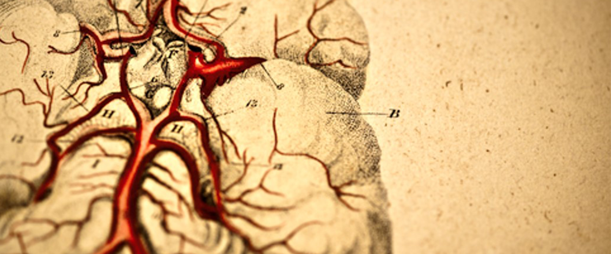
BRAIN ANEURISM
BRAIN ANEURISM
Aneurysm is a ballooning in the vessel due to weakness in the vessel wall. These weak areas may include the entire wall of the vessel (fusiform), outward swelling may occur only from a section of the wall (sacular) or may separate the vessel wall layers (dissecting). Aneurysms can occur in all veins in the body. When aneurysms in the brain vessels burst, they can cause serious brain damage and death.
Aneurysms can be seen in 6 out of every 100 people in the society. Generally, they are more common in women. More than one aneurysm can be seen at the same time in one fifth of the patients. The most common ages are between the ages of 40-60. The most important risk factors are smoking and family history of aneurysm.
How is aneurysm diagnosed?
Unfortunately, the majority of aneurysms are not detected until they burst and cause cerebral hemorrhage. Sometimes large aneurysms can cause symptoms such as visual disturbances, pain behind the eyes, headache, and nerve palsy before they burst. Since brain magnetic resonance imaging (MRI) and brain tomography are performed more often than before, these aneurysms can be detected more frequently without bursting. MRI and CT show the presence of aneurysm, but cerebral angiography is required for detailed evaluation of the aneurysm and treatment planning. Angiogram is obtained by placing a thin flexible tube called catheter from the groin area into the artery to visualize the brain and neck vessels and then advancing it up to the neck vessels and injecting a liquid that makes the vessels visible under X-rays. Detailed images of the veins at different angles are obtained and the relevant veins are evaluated. Features such as size, shape and location of the aneurysm are evaluated and this information is used to select the most appropriate treatment methods and techniques.
What happens if the aneurysm bursts (ruptures)?
When the aneurysm bursts, the blood fills the space between the meninges (subarachnoid hemorrhage) and causes severe headache. Patients sometimes describe this headache as "the worst headache of their life" or "there was a bomb or lightning in my head". Other symptoms are nausea, vomiting, impaired consciousness, weakness or drowsiness. Subarachnoid hemorrhage is an emergency, and about one-fifth of patients die before they can be delivered to the hospital. 40-50% of the patients who reach the hospital die within the first 30 days. Half of the surviving patients are disabled if they do not receive the necessary treatment.
Patients with aneurysm-related cerebral hemorrhage stay in the intensive care unit for up to 2 weeks. After bleeding, swelling and fluid accumulation in the brain cavities called hydrocephalus can be seen. This may cause increased intracranial pressure and may require a drain tube to be inserted into these spaces to reduce pressure. It can also cause contraction and contraction of blood vessels in the brain cavities (vasospasm). Since vasospasm will reduce the amount of blood reaching the brain, these patients may experience stroke. In order to prevent this situation, vasodilator drugs are given to the patients in intensive care and their blood pressure is kept at high levels. If these treatments are not successful, patients may need to undergo brain angiography again, and vasodilator drug injected directly into the blood vessels or dilated with a balloon.
How are aneurysms treated?
If the aneurysm has caused a brain hemorrhage, it must be treated. Not all aneurysms that are detected without causing bleeding need to be treated. Aneurysms that are very small in size and detected incidentally can be followed up with close observation without treatment. Experts in this field provide recommendations to the patient on treatment based on the size and location of the aneurysm and the patient's history. There are two options for treatment: Open surgery, ie clipping and closed method, also known as the endovascular method.
Open Surgery
In the open method, after the skin is cut and the skull is opened by neurosurgeons, a metal clip is placed in the aneurysm neck after reaching the brain area where the aneurysm is located. Thus, blood flow is prevented from entering the aneurysm.
Closed-Endovascular Method
In the closed method, interventional neuroradiologists approach the cerebral angiography from the inguinal vein similar to the cerebral angiography, reaching the neck and brain vessels with the help of very thin catheters, respectively, and exclude the aneurysm from the bloodstream with various techniques. The most common of these methods is filling the aneurysm with platinum coils called coils. Stent and balloon can also be used to assist the coiling process. In another method, without entering the aneurysm, a special stent called a flow transducer is placed into the vein where the aneurysm is located, and the blood flow is removed from the aneurysm and directed to the normal circulation. Thus, the pressure within the aneurysm decreases and the aneurysm spontaneously collapses over time. The most appropriate of these techniques is selected according to the size, shape, location of aneurysms and their relationship with neighboring vessels. Patients to be treated with a stent should use blood thinners before the procedure.
Although both methods are effective, the closed method is currently used as the first option in the majority of aneurysms. The fact that it is less risky and more comfortable for patients compared to open surgery has brought the closed method to the fore all over the world. The most extensive studies have shown that there is less disability and death after treatment with the closed (endovascular) method compared to open surgery. At the same time, less epilepsy and decreased cognitive abilities were observed in patients who were applied the closed method.