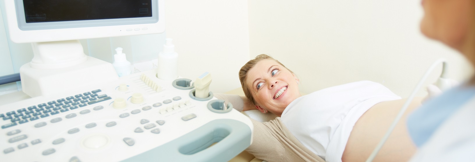
Perinatology
Tınaztepe Health Group Maternal-Fetal Medicine Unit provides globally competitive, reliable and qualified service with experienced expert staff to protect the health of our patients, examine, analyze, diagnose and treat existing diseases.
What are High-Risk Pregnancies?
If you have a chronic disease such as diabetes, goiter, high blood pressure before you become pregnant, you have a pregnancy under the age of 18 or over 35, if you are overweight or overweight, you have a risky pregnancy if you take medication. Sometimes these problems can arise with the effect of pregnancy. You may experience bleeding during pregnancy. All these situations put you in the risky pregnancy category. If you have had any problems with previous pregnancies (premature birth, the early arrival of water, placenta previa, ablation placenta, pregnancy poisoning), your current pregnancy is also considered in the risky pregnancy category.
Malformation Ultrasonography: Detailed ultrasound, level II ultrasound, color ultrasound, and four-dimensional ultrasound among the public names such as the examination of pregnancy 18-22. Ultrasonographic examination. In this examination, structures such as "head, brain, face, neck, thorax, heart, large vessels, abdominal wall, stomach, kidneys, bladder, spine, skeleton, arms, hands, legs, feet" of the baby are evaluated systematically in terms of anomaly findings.
Shortness or absence of nasal bone in baby, accumulation of fluid in the nuchal pleura, presence of cyst in the brain (choroid plexus cyst), fluid expansion in areas where cerebrospinal fluid circulates (ventriculomegaly), shortening of arm and thigh bones, urinary collections of the kidney (pyelectasis), the presence of chromosomal abnormalities such as having a single umbilical artery, ventricular septal defect in the heart, rocker bottom in the feet, and club foot are evaluated.
Perinatology Unit Procedures:
CVS (Chorionic villus sampling, placenta removal): It is a method in which the placental tissues in this region are collected with a syringe applied with negative pressure by means of back and forth movements by entering the placenta (the last organ which feeds baby) with the needle of the appropriate size. It is done between 10th-13th weeks of pregnancy. There is a 1% risk that the baby will die in the womb.
Amniocentesis: Ultrasonography is the procedure of taking a 15-20 cc sample from amniotic fluid that the baby swims in by entering the mother's abdomen with the needle of appropriate size. Some of the diseases of the baby (such as Down syndrome) can be diagnosed by the production of spilled cells from the baby in this fluid by special methods. This process is carried out in the 15th-20th weeks of pregnancy. There is a risk of pregnancy loss of 1/250 after the procedure.
Cordocentesis It is the procedure of taking blood from the veins in the umbilical cord of the baby by entering the mother's abdomen with an appropriate size needle accompanied by ultrasonography. This blood sample can be used to diagnose certain infant diseases (such as Down's syndrome). This is done after the 20th week of pregnancy. There is a 1-2% risk of pregnancy loss after the procedure.
Intrauterine blood transfusion: Infants who develop severe anemia in the womb (Hydrops Fetalis), such as the appropriate amount of blood cordocentesis process, the umbilical cord or infant's abdominal membrane is tried to correct the anemia.
The Amnioinfusion: In some special cases, it is the process of introducing fluid to the infant in which the baby is swimming, by entering the needle with the needle as in amniocentesis.
Amnioreduction: In some special cases, the excess amount of fluid that the baby floats in is gradually discharged to the outside with the needle of the appropriate size, since it reaches a very large amount of water in the womb and creates a risk of premature birth.
Fetal Bladder Urine sampling: In some special cases, as in amniocentesis, in order to evaluate the renal functions of the baby, it is the procedure of entering the baby's urine bag with a needle from the abdomen and taking a fetal urine sample.


